


3.) Protein Story Phase:
CRISPR-Cas9
Climbing the Mountain
CRISPR is a naturally occurring bacterial and archaeal immune system being engineered for biotechnology applications.
The resources below will introduce the basics of the CRISPR-Cas9 protein. You will then choose a specific protein story related to Cas9 that you want to model. You will read a scientific research article, find a Cas9 protein structure file, and design a 3D printed physical model of a Cas9 protein.
Note: If you skipped the Training Phase and need to review basic protein structure-function concepts, visit the Training Phase Protein Structure Resources.
Recommended Models for the CRISPR-Cas9 Protein Story Phase
The MAPS program revolves around using physical models as teaching tools. So if at all possible, we encourage you to use the recommeneded models below for the Cas9 protein story phase.
All recommended models are available for purchase through 3D Molecular Designs. Use the discount code below to receive a 10% MAPS discount on your 3D Molecular Designs purchases. Some models are available to borrow free of charge through the MSOE Lending Library.

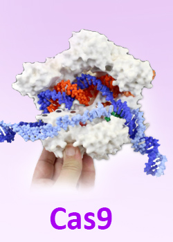
1. Introduction to CRISPR-Cas9
An Introduction to CRISPR-Cas9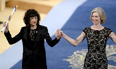
You have probably been hearing a lot about CRISPR lately. It is a powerful technology that has been revolutionizing the way we do research and it is becoming a viable option for treating many genetic disorders. The possiblilties for protein engineering Cas9 for specific applications are almost endless.
You might be aware that Jennifer Doudna and Emmanauelle Charpentier were awarded the Nobel Prize for Chemistry for their research on solving the Cas9 structure and elucidating its function based on the structure.
2. The Flow of Genetic Information and Non-Coding RNA
"This overall flow of genetic information - from DNA to RNA to protein - is known as the central dogma of molecular biology, and it is the language used to communicate and express life."
- Jennifer A. Doudna, A Crack in Creation
In this section we are going to explore the flow of genetic information, from DNA to RNA to Protein, along with non-coding RNA. These key concepts are crucial to understanding how the CRISPR-Cas9 protein functions.
Note: If you skipped the Training Phase and need to review basic protein structure-function concepts, visit the Training Phase Protein Structure Resources.
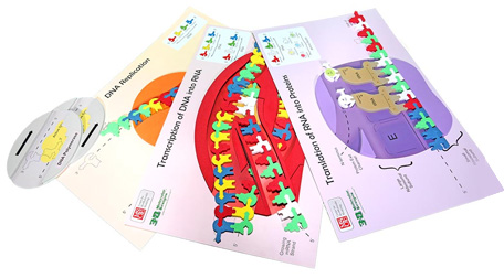
The Flow of Genetic InformationUnlike most MAPS proteins, Cas9 is unique because it is a protein that needs RNA to function! How does that work? Well first you will need to understand the flow of genetic information from DNA to RNA to protein. Then we will explore some fun exceptions to the rule!
The Flow of Genetic Information Kit from 3D Molecular Designs is an excellent modeling tool for learning the basics of DNA replication, RNA transcription and protein translation.
Replication Activities
Transcription Activities
Translation Activities
3. The Guide RNA
"...how a piece of RNA could pair up with its phage DNA counterpart and cause that DNA to be destroyed."
- Jennifer A. Doudna, A Crack in Creation
Not all RNA is destined to be translated into protein. Some is ready to provide function as RNA. We call these RNAs non-coding RNAs. You are probably familiar with transfer RNA (tRNA) and ribosomal RNA (rRNA), which are non-coding RNAs. But there is a wide range of non-coding RNAs with in the human genome with myriad functions that researchers are still trying to understand. From long non-coding RNAs, to microRNA, snRNA, siRNA and many others, this is a hot area of research and there is so much more to be learned!
The Complementarity Activity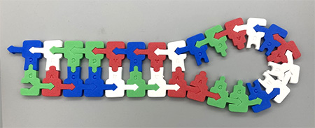
In order for RNA to have intrinsic function, like proteins, they also fold into 3D shapes like proteins. However, their structure is not based on the amino acid sequence, it is based on the nucleottide sequence. Stretches of non-coding RNA have areas of complementarity that allow then to fold over and bind to themselves in a sequence-dependent fashion. In order to model how this works, do the following activity and then come back for more discussion.
Click here to download the Complementarity Activity (PDF)
Post-activity discussion
Hopefully you discovered that complementary sequences in RNA can cause it to fold onto itself and base-pair to itself. this is called a hairpin turn (looks a little like a bobby pin, right?) forming what is known as a stem-loop structure, with the base-paired section being the stem, and the nucleotides in the middle being the loop. We will see this structure in the non-coding RNA involved in Cas9 function later. When you opened your RNA up and "reverse-transcribed" it to DNA sequence, what did you find that was unique about the sequence on the 2 strands? Hopefully you saw that the complementary sequences on one RNA strand are transcribed from identical sequences on opposite strands of DNA. We call this a palindrome, like "radar" or "racecar", or "Sit on a potato pan, Otis". Ok, I couldn't resist that last one lol! Anyway, in DNA, the palindrome you created was found on complementary strands, reading 5' to 3'. We will find palindromes later in this module and can discuss their meaning further then.
In addition to forming hairpin turns and step loop structures, RNA also twists into 3-D structures due to a 'stickiness' of its backbone caused by the 2'OH group (missing in deoxy-ribonucleic acid) that allows it to hydrogen bond to other parts of the RNA molecule by acting as a hydrogen bond donor. No need to understand this deeply unless you want to dig deeper. This is just to explain that non-coding RNA folds into unique 3D structures based on complementarity and a sticky backbone!
Forming the Guide RNA and the Cas9 Surveillance Complex
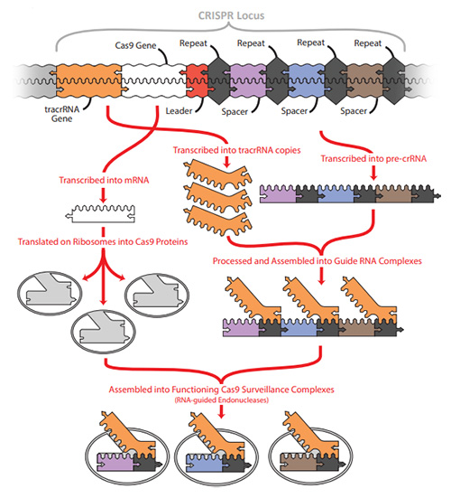
For this module we are focused on non-coding RNA in the CRISPR system. There is one section of RNA that is transcribed from sequence inserted from a previous viral attack (known as spacer sequence) and its adjacent repeat sequence, together known as CRISPR RNA (crRNA). There is another section of RNA that is transcribed upstream of the CRISPR array (where all the viral sequences are kept, separated by repeats), called the trans-activating RNA or tracrRNA.
The tracrRNA has a section that is complementary to the repeat section attached to each spacer sequence. This allows it to bind to each spacer sequence in the CRISPR array by base-pairing with the attached repeat (no matter what the unique spacer sequence is - brilliant right?). These are cleaved so that dual-guide RNAs are formed, made up of a tracrRNA base-paired to a repeat with an adjacent spacer. In this way, guide RNAs can be formed that are complementary to every viral sequence in the CRISPR array of that bacterium. Cas9 “grabs” these guide RNAs which changes the conformation of Cas9 to convert from an inactive protein into an active Cas9 surveillance complex.
These surveillance complexes hang out in the bacterium, searching for complementary sequences to their spacer sequences. They do this by binding DNA at PAM (proto-spacer adjacent) sequences. When the spacer sequences are being excised from the attacking virus and inserted into the CRISPR array of the bacterium, it is done in a specific manner. It finds a PAM sequence, which for Cas9, is 5’ N-G-G, where N can be any nucleotide. This simple sequence is found multiple times in any genome. However, if the segment of the viral genome that is cleaved (protospacer) is adjacent to this sequence, it can act like a bookmark to help it easily find that sequence in future invading viruses. In addition, it helps the bacterium to distinguish the invading virus sequence from its own CRISPR array sequence because the PAM sequence (5’ NGG adjacent to the protospacer) is only found in the virus. Once the sequence is inserted into the bacterial CRISPR array, it is then known as a spacer (no longer a protospacer) and has no PAM sequence associated with it.
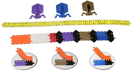
The CRISPR Adaptive Immunity Kit
The CRISPR Adaptive Immunity Kit is an excellent modeling tool for exploring the CRISPR adaptive immunity system and the formation of the Guide RNA and Cas9 Surveillance Complex. Work through the Adaptive Immunity Kit if you have access to one and then watch the video summary below.
CRISPR Adaptive Immunity Kit
Review of the CRISPR Adaptive Immunity Kit
4. CRISPR-Cas9 Structure and Function
"...how a piece of RNA could pair up with its phage DNA counterpart and cause that DNA to be destroyed."
- Jennifer A. Doudna, A Crack in Creation
Making the Cut with CRISPR-Cas9
Now we have a good understanding of how the bacterium arms itself with Cas9 surveilance complexes that each contain guide RNA with sequences complementary to a previous viral attack. Now let's explore how those complexes seek out and destroy incoming bacteriophage!
Use the Making the Cut kit to model how this works and then watch the video below, which well help summarize what you have been exporing up to this point.
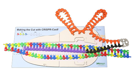
Making the Cut with CRISPR-Cas9
Review of the Making the Cut with CRISPR-Cas9 Kit
So now we have a big picture idea of how Cas9 binds PAM sites in DNA that enters the bacterium, checks the adjacent sequence to see if it matches the spacer sequence in the guide RNA, and makes a double-stranded cut in the DNA if there is a match! Go back and work with the Making the Cut Kit again if you want to clarify anything before moving on.
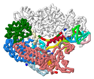
As mentioned, Cas9 is a fascinating protein because of its use of guide RNA, and because of its multiple functional domains. Functional domains are specific parts of the protein that do specific jobs. In some CRISPR systems, many different proteins form a complex and each do various jobs. Diffferent classes of CRISPR systems have evolved so that some, like Cas9, have all those jobs packed in one protein! Functional domains have specific structure-function characteristics and can often be found in many proteins. For this reason, once they are well characterized in one protein, they are named the same when they are found in other proteins.
For example, the 2 endonuclease domains in Cas9 (the parts of Cas 9 that cut the target and non-target strands of DNA) are called HNH, and RuvC, respectively. Because their sequences are homologous to other endonuclease domains discovered in other proteins, it gives us great insight into how they are likely to function in Cas9. Researchers know that each of the active sites have an invariable amino acid that, when mutated, will inactivate the endonuclease activity. This information allows researchers to engineer Cas9 so that it is a nickase, that is a protein that will cause a single-stranded nick in either the target or non-target strand (depending on which domain is mutated) rather than a blunt, double stranded cut that normally occurs. Why would this be helpful? Discuss your thoughts and stay tuned for the section on Cas9 technology applications.
Domain Activity
The resources below will help you to explore the functional domains of Cas9 and their structure-function relationships. The video below will help guide you as you explore the domain model on paper and/or within Jmol. Stop the video and take notes as you go. Discuss with your colleagues. There is a lot of info so take your time.
- Printable domain graphic (11x17 PDF)
- Printable domain graphic (8.5x11 PDF)
- CRISPR Domains Jmol Visualization
The Functional Domains of CRISPR-Cas9
Additional Resources
- "CRISPR-Cas9 Sturctures and Mechanisms" by Jiang & Doudna (PDF) - most of the information in the video above came from this review. You might also want to go back and use the Making the Cut Kit again now that you have explored the domains as it might make more sense to you now or it might make you think of it in a different way. Multiple different models can often help us think about things in a deeper way and clarify confusing concepts.
- Cas9 Functional Domains Overview (PDF)
- Cas9 Domains Jmol Exploration (webpage)
- Labeling the Cas9 Domains Interactive Digital Activity - Version 1 (Powerpoint)
- Labeling the Cas9 Domains Interactive Digital Activity - Version 2 (Powerpoint)
Review of the Functional Domains of CRISPR-Cas9
5. Defining Your CRISPR-Cas9 Protein Story
"The more we know, the more we realize there is to know"
- Jennifer Doudna
Now that you are armed with an extensive understanding of what Cas9 does and how its structure is important to its function, it is time to decide what specific CRISPR-Cas9 story you would like to explore.
The expandable content below will give you some suggested topics you may choose to focus on, recommended Cas9 research papers and reviews to read, and link to a collection of Cas9 structure files to use when designing your 3D printed protein model.
Cas9 Genome Editing TechnologiesWhat Does it Mean to "Design" a Protein Model?
Tim Herman, PhD Explains How to Approach Your Model Design in Jmol
"Designing" a protein model means you explore a protein structure in Jmol, and then simplify the way the protein is visually displayed to make the key features of the protein that help communicate your molecular story more obvious.
This can mean hiding some atoms that are not important for your protein story, changing the display format of certain parts of your protein structure or changing colors to best highlight the most important parts of the protein structures. In the next section, you will learn the Jmol commands needed to accomplish this.
How to Approach Your Model Design
Tim Herman, PhD Explains How to Approach Your Model Design in Jmol
All 3D printed protein models made in the MAPS program are designed using the program Jmol. You start with a protein structure file (.PDB file), which contains the 3D locations of all of the atoms that make up the protein. Using the Jmol commands you will learn below, you can then edit the way the protein is displayed, hiding some atoms, changing display formats and customizing colors. When happy with your design, you can export your protein model from Jmol in file formats suitable for 3D printing.
We have created a detailed Jmol Training Guide that will cover everything you need to know to design and build your model. The Jmol Training Guide is broken down into four main sections, which we strongly recommend you explore in order until you are comfortable with Jmol and designing protein models for 3D printing.
- 1) Getting Started:
- Accessing Jmol
- Loading Structures
- Saving and Reloading - 2) Simplifying Your Protein:
- Exploring the Structure
- Focusing on the Backbone - 3) Adding the Details:
- Adding Sidechains
- Adding Ligands
- Adding Molecular Bonds - 4) 3D Printing Your Design:
- Adding Support Struts
- Checking Your Design
- Print it Yourself
- Pay Someone Else to Print it
Moving to the Next Phase
Once your team has finished the protein story phase, move on the Capstone Experience, where you will learn how to present your project using your 3D printed model and a variety of complimentary digital and print media.