Building Your Physical Protein Models
The final step to successfully competing in the Science Olympiad Protein Modeling Event is to actually build your protein models. This section will cover how to construct a protein model, including information on:
- The visualization environment
- Folding a Mini-Toober model
- Taking your model further and judging tips
The Visualization Environment
Overall Layout of the Visualization Environment
Changing the Display Format and Color Schemes
Using the Select and Restrict Commands
Advanced Jmol Training Guide
For students that really want to push their understanding and knowledge of Jmol further, the advanced Jmol training web pages below will provide a more detailed review of commands and techniques for manipulating protein and DNA structures in the Jmol visualization environment.
- Getting Started in Jmol
- Finding Protein and Molecular Structures
- Changing Colors and the Display Format
- Saving and Reloading Your Work
- The Select Command and Boolean Operators
- Adding Sidechains to Your Structure
- Bonds and Structural Supports
- Working with Nucleic Acids
Folding a Mini-Toober Model
Every team competing in the Science Olympiad Protein Modeling Event will be expected to create a pre-build protein model of specific protein structure. The pre-build model will be created using a purchased "pre-build" Mini-Toober kit from www.3dmoleculardesigns.com, or with found materials of the participants' choosing such as KwikTwist tie-down ropes.
The same pre-build model will be used at each level of the competition (invitational, regional, state and national). Participants will take their pre-build model home after each event.
The pre-build model must be impounded prior to the beginning of the event (usually the morning of the event. The pre-build model should be submitted with a 4"x6" card with a written description of the model's colors and creative additions, addressing what, how, and why. Check the official rules for details
This section contains three important video tutorials describing how to map out and fold these models.
Mapping Out the Mini-Toober Model
View video content as a PDFFolding Secondary Structures
View video content as a PDFFolding the 3-D Shape
View video content as a PDFA Note on the Length of Your Toober
An astute student may notice that the length of the Toober included in a prebuild modeling kit purchased from 3D Molecular Designs appears to be one amino acid length (2cm) too short. For example, if you are modeling amino acids 1-85, you would expect the length of Toober to be 170cm long (at a scale of 1 amino acid = 2cm) - but it is actually 168cm long.
The video below attempts to explain this apparent discrepancy using schematic graphics and Jmol renderings.
View Graphic as a PDF
Taking your model further and judging tips
Pre-build models are expected to have additional added features and all models will undergo a judging process. This section contains two important video tutorials describing how to approach adding additional features on the pre-build model and how models will be judged.
Going Beyond Just the Backbone
View video content as a PDFHow do you know which residues are important?
The protein that you are modeling may be large and complex. Here are a few simple ways to figure out which amino acid residues in the structure are important for its structure or function:
- Are there specific residues mentioned/discussed in the abstract and/or manuscript describing this protein structure? Here are some possibilities:
- These amino acids may be mentioned as the active site, catalytic, or binding site residues.
- They may also be discussed as sites of mutations that change the structure and/or function of the protein.
- Are there any ligands, drugs, ions, inhibitors or other small molecules bound to the protein structure?
- The residues within ~5 angstrom of the small molecule are likely to be non-covalently interacting with it. These amino acid residues may have an important role in the structure and function of the protein.
- Any residues covalently linked to the small molecule ligands, drugs etc. may be critical to the structure and function of the protein.
- Does your protein of interest interact with other proteins, nucleic acids?
- Look through the figures in the manuscript describing the protein structure. Pay attention to any additional proteins, DNA, RNA or other molecules shown in these figures.
- Read about these molecules and try to figure out how and why the interaction is shown.
- Can you figure out the interactions stabilizing these molecules?
- Examine residues at the interface between 2 proteins or a protein and DNA/RNA etc.
- These amino acids may have important roles in the functioning of the protein.
Judging the Protein Models
View video content as a PDF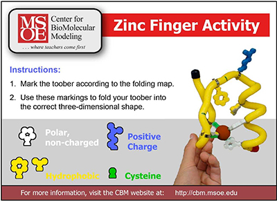
Zinc Finger Example Model
The CBM's Zinc Finger Folding Kit works as an example modeling activity for the Science Olympiad Protein Modeling Event. The model is available to borrow free from the CBM Lending Library, and can be used alongside the online Zinc Finger Design Environment.
Also available are example Scoring Rubrics and Scoring Spreadsheets for this zinc finger activity, which mimic the rubrics and scoresheets used by event supervisors in the Science Olympiad Protein Modeling Event.
Exploring the RCSB Protein Databank for the Onsite Jmol Exploration
This year, instead of modeling a protein during the on-site competition, teams will answer questions as part of a "Jmol Exploration".
The purpose of the Jmol exploration component of this year’s event is to allow students to demonstrate their skill in using Jmol to manipulate and explore a pdb file, and navigate the RCSB Protein Data Bank website (www.rcsb.org).
The video below will describe the RCSB Protein Databank Website, and the features teams should be familiar with for the onsite "Jmol Exploration" portion of the competition.