The 2016 Science Olympiad Protein Modeling Event
Proteins Involved in the Biosynthesis and Subsequent Signaling of the Neurotransmitters Dopamine and Serotonin
Inspiration for the theme: Sepiapterin Reductase and the Beery Twins
 Be extra sure to explore the information in all three tabs below
Introduction
In the video below, Tim Herman, Ph.D. from the MSOE Center for BioMolecular Modeling provides an introduction to this year's topic, Sepiapterin Reductase and the Beery Twins.
The Beery family story is a genomics story. It is an example of how whole genome sequencing was used to arrive at a definitive, molecular diagnosis of the twins’ medical condition.
Your primary resource in exploring this story will be the original research paper published by the research group at the Baylor College of Medicine who sequenced the twins’ genome and then identified mutations in the sepiapterin reductase gene as the cause of their dystonia disorder. A copy of that paper is provided here:
Paper Abstract:
Whole-genome sequencing of patient DNA can facilitate diagnosis of a disease, but its potential for guiding treatment has been under-realized. We interrogated the complete genome sequences of a 14-year-old fraternal twin pair diagnosed with dopa (3,4-dihydroxyphenylalanine)–responsive dystonia (DRD; Mendelian Inheritance in Man #128230). DRD is a genetically heterogeneous and clinically complex movement disorder that is usually treated with L-dopa, a precursor of the neurotransmitter dopamine. Whole-genome sequencing identified compound heterozygous mutations in the SPR gene encoding sepiapterin reductase. Disruption of SPR causes a decrease in tetrahydrobiopterin, a cofactor required for the hydroxylase enzymes that synthesize the neurotransmitters dopamine and serotonin. Supplementation of L-dopa therapy with 5-hydroxytryptophan, a serotonin precursor, resulted in clinical improvements in both twins.
Many of the figures from the Lupski and Gibbs paper are discussed below, along with other resources and background material you will need as you explore the Beery family story.
Table of Contents:
- The Beery Family
- The Beery Family History
- The Beery Family Pedigree
- The Genetic Basis of the Beery Twins' DOPA-Responsive Dystonia
- Genome Sequencing of the Beery Twins
- Validation of Two Sepiapterin Reductase Alleles by Sanger DNA Sequencing
- The Full Sepiapterin Reductase Gene
- The Role of Sepiapterin Reductase in
the Biosynthesis of Two Neurotransmitters
- Neurons and the Synaptic Cleft
- Sepiapterin Reductase - What Does It Do?
- What does the cofactor BH4 have to do with the Beery family story?
- A Summary of Serotonin and Dopamine Biosynthesis
- Mapping the Mutations to a
Physical Model of Sepiapterin Reductase
- The Robert Huber Paper
- The Locations of Retta and Joe Beery's Mutations
- The Solution
- But Life is Complicated
- The Beery Family Today
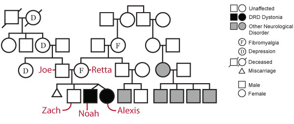
The Beery Family
The Beery Family History

The Beery Twins, Noah
and Alexis, as Children
Soon after Alexis and Noah Beery were born it was obvious to their parents, Joe and Retta, that the twins were different from their older brother, Zach. They demonstrated poor muscle tone, cried nonstop, vomited frequently and missed developmental milestones. Physicians diagnosed the twins with cerebral palsy. By age 5, Alexis was having difficulty swallowing and was wasting away, symptoms not consistent with cerebral palsy. Retta came across an article about a rare disorder, dopa-responsive dystonia (DRD), which is caused by a deficiency of the brain neurotransmitter dopamine. The symptoms matched those of her daughter. Shortly after, both twins were treated with L-DOPA (dopamine precursor), and their condition improved dramatically. However, as the twins grew older, it became apparent that even this diagnosis was incorrect as their health began to further deteriorate.
To learn more about the Beery family and their journy, explore their website at http://dystonia.thebeerys.com.
The Beery Family Pedigree
Examination of the Beery family pedigree shows a history of depression on the paternal side and a history of fibromyalgia and undisclosed neurological disorder on the maternal side, in addition to DRD in the twins.
The Genetic Basis of the Beery Twins'
DOPA-Responsive Dystonia
Genome Sequencing of the Beery Twins

An Example of Genome Sequencing Data
In the case of the Beery twins, whole genome sequencing led to a more complete understanding of the molecular basis of their disease and informed a change in their medical treatment. Joe Beery, Alexis and Noah's father, was working as the VP for Information Technology at Life Technologies (a biotech company involved in NextGen DNA sequencing) and arranged to have the twins' genomes sequenced by a group at the Baylor College of Medicine. More than 2 million variations were found in the DNA sequence of the twins' genomes, compared to a reference human genome. This level of variation is quite normal. Various bioinformatics filters were applied to eliminate variants that were not thought to be responsible for their condition. Following this filtering, only three variants remained as candidate genes. One of these was sepiapterin reductase (SPR). The nucleotide sequence of the twins' SPR genes revealed two different mutations – each of which were believed to result in an inactive enzyme.
Validation of Two Sepiapterin Reductase Alleles by Sanger DNA Sequencing
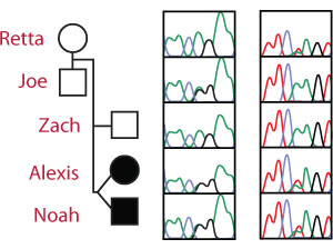
The SPR Genotype Sequencing Traces
Recent advances in DNA sequencing technology have made it possible to rapidly sequence whole human genomes at a cost approaching $1,000. Although NextGen DNA sequencing is rapid and inexpensive, it is not as accurate as other sequencing methods. Therefore, Sanger DNA sequencing was used to confirm the sequence of the twin’s SPR genes.
The Sanger sequencing traces (right) show the SPR genotype for each member of the Beery family.
The Arg150Gly mutation is an A → G mutation on chromosome 2 at nucleotide 72,969,094 leading to the replacement of Arginine with Glycine. The unaffected father is heterozygous (A/G) for the pathogenic Arg150Gly allele at the first locus and homozygous (A/A) for the wild-type allele at the second locus.
The Lys251X mutation is an A → T mutation on chromosome 2 at nucleotide 72,972,139 resulting in the conversion of a Lysine codon (AAG) to a STOP codon (UAG). The unaffected mother is homozygous (A/A) for the wild-type allele at the first locus but heterozygous (A/T) for the pathogenic Lys251X allele at the second locus.
Each affected twin is a compound heterozygote - (A/G and A/T) with a different pathogenic mutation on each allele.
The Full Sepiapterin Reductase Gene
Explore the sepiapterin reductase gene map shown below to see Retta Beery's mutation (highlihgted in pink) and Joe Beery's mutation (highlihgted in blue). Click Here to download the entire sepiapterin reductase gene as a PDF document.

The Role of Sepiapterin Reductase in
the Biosynthesis of Two Neurotransmitters
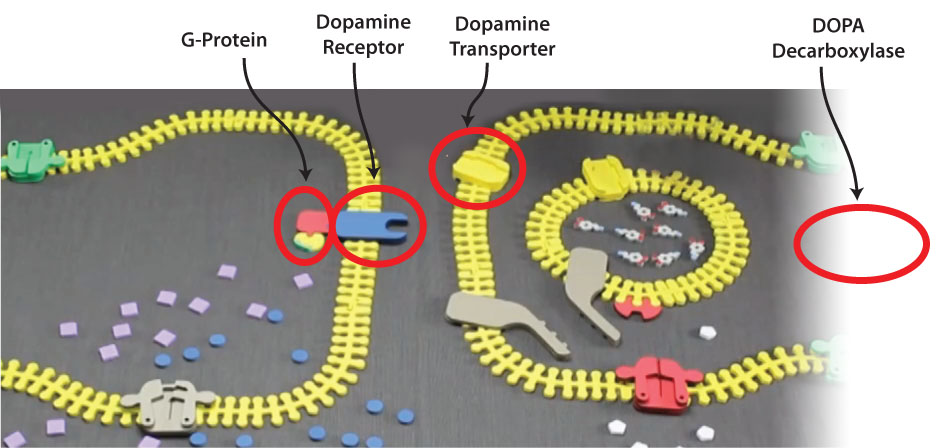
Neurons and the Synaptic Cleft
Sepiapterin reductase is an enzyme associated with the biosynthesis of two neurotransmitters, dopamine and serotonin. But to understand the specific role of sepiapterin reductase in our nervous system, an overview of neuron structure and the synaptic cleft is needed. In the two videos below, Gina Vogt from the MSOE Center for BioMolecular Modeling discusses the shape of neurons, the location of the synaptic cleft and the role of dopamine and other neurotransmitters in the neurotransmission.
Sepiapterin Reductase - What Does it Do?
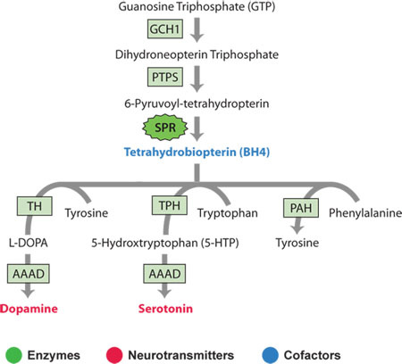
Sepiapterin reductase is an enzyme that is responsible for the biosynthesis of a cofactor known as tetrahydrobiopterin (BH4).
Enzyme cofactors are small molecules that enable some enzymes to catalyze difficult reactions that would otherwise be impossible with only the twenty amino acid sidechains. In other words. . . most enzymes contain "active sites" that bind specific substrates in such a way that several active site amino acid sidechains are precisely positioned to perform a chemical reaction. Sometimes these active site sidechains require the help of cofactors to create the necessary chemistry to catalyze the reaction.
What does the cofactor BH4 have to do with the Beery family story?
The cofactor BH4 is needed by an enzyme that participates in the biosynthetic pathway that converts the amino acid tyrosine into the neurotransmitter dopamine. A similar enzyme uses BH4 to convert tryptophan into serotonin. Both of these enzymes are "hydroxylases" that add a hydroxyl group to an amino acid.
In the video below, Tim Herman, Ph.D. from the MSOE Center for BioMolecular Modeling provides an overview of neurotransmitter biosynthesis and the role sepiapterin reductase plays in the pathway.
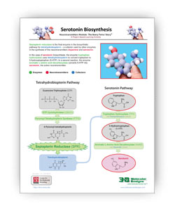
A Summary of Serotonin and Dopamine Biosynthesis
The two .PDF links below summarize the steps needed to turn tryptophan into serotonin and tyrosine into dopamine.
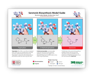
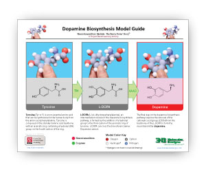
Sepiapterin reductase is the final enzyme in the biosynthetic pathway for tetrahydrobiopterin – a cofactor used by other enzymes in the synthesis of the neurotransmitters dopamine and serotonin.
In the case of dopamine biosynthesis, the enzyme tyrosine hydroxylase uses tetrahydrobiopterin as a cofactor to convert tyrosine to L-DOPA. In the case of serotonin, the enzyme tryptophan hydroxylase uses tetrahydrobiopterin to convert tryptophan to 5-hydroxy tryptophan (5-HTP). In a second reaction in each pathway, aromatic L-amino acid decarboxylase (AAAD) converts both L-DOPA to dopamine and 5-HTP to serotonin, the active neurotransmitters.
Click here for expanded definitions of Enzymes, Neurotransmitters and Cofactors
The two .PDF links below summarize the two pathways involved in producing tetrahydrobiopterin and neurotransmitters.

Mapping the Mutations to a
Physical Model of Sepiapterin Reductase
The Robert Huber Paper
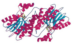
The 3-dimensional structure of a mouse sepiapterin reductase was first determined by Robert Huber’s group in 1997. An annotated version of the "primary citation" (the original research paper that reported this structure) is provided below. This is a very detailed research paper. You are not expected to read and understand the entire paper. To guide your reading, we have highlighted those sections of the paper that will provide you with a general introduction the structure and function of the protein.
Note that your pre-build model is based on the structure of a mouse sepiapterin reductase protein, as described by 1sep.pdb. We chose the mouse enzyme for this protein modeling event because:
- It was the first structure of this protein that was solved (in 1999).
- Tetrahydrobiopterin was bound in the active site of this enzyme.
- The mouse enzyme is 74% identical (and 88% similar) to the human enzyme.
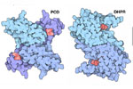
The Locations of Retta and Joe Beery's Mutations
As you prepare for this protein modeling event, you should determine the molecular consequence of the two mutations identified in the twins’ SRP gene – and map these changes onto the 3D structure of the sepiapterin protein.
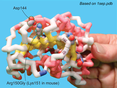
Joe Beery's Mutation, an Arginine to a Glycine
Joe Beery’s Arg150Gly missense mutation (left image) results in a change from a positively charged basic arginine amino acid at position 150 to an uncharged glycine. This would disrupt a salt bridge interaction with the negatively charged aspartic acid at 144 in the functional enzyme.
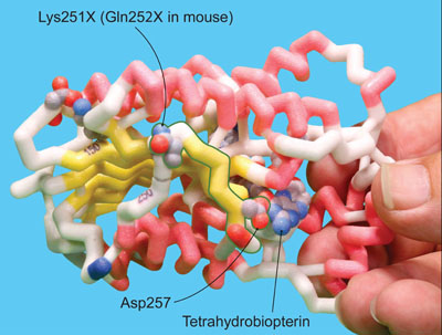
Retta Beery's Mutation, a Lysine to a STOP Codon
Retta Beery’s Lys251X nonsense mutation (right image) results in a change from lysine at position 251 to a premature STOP codon, causing truncation of the enzyme.
The Solution
If Alexis and Noah can’t make enough BH4 to support normal levels of dopamine and serotonin, how can we fix this problem?
The solution is to provide the neurotransmitter precursor immediately downstream from the biosynthetic step that requires BH4 – namely L-DOPA and 5-hydroxy tryptophan. Daily doses of these two precursors allow Alexis and Noah to lead the lives of normal, healthy teenagers.
But Life is Complicated
As one final twist in this story, the L-DOPA and 5-hyroxy tryptophan medication that Alexis and Noah take is also supplemented with another small molecule called carbidopa. Can you figure out what carbidopa is and what role it plays in the twins' medication?
The Beery Family Today

The Beery Twins Today
Noah and Alexis have a passion to share their story. According to a recent blog post, "It's our hope and prayer that people will use their voices in their medical care or any other area of their lives, and never, ever give up hope - no matter how dire the situation may seem. Because hope is unlimited and has no boundaries!"
In order to truly appreciate and successfully model this year's Protein Modeling Event structures, a thorough understanding of proteins, DNA and genomes is needed. This section will explore these amazing macromolecules in more detail using suggested physical model kits, online resources and additional websites.
Protein Structure
Proteins are long linear sequences of amino acids
that fold into complex 3-dimensional shapes
following basic principles of chemsitry.
In this first of two videos on protein folding, Tim Herman, Ph.D. from the MSOE Center for BioMolecular Modeling uses the Water Cup from 3D Molecular Designs to demonstrate some basic principles of chemistry.
In this second of two videos on protein folding, Tim Herman, Ph.D. from the MSOE Center for BioMolecular Modeling uses the Amino Acid Starter Kit from 3D Molecular Designs to demonstrate how the basic principles of chemistry directly affect protein folding.
In this video, Tim Herman, Ph.D. from the MSOE Center for BioMolecular Modeling uses the Alpha-helix Beta-sheet Construction Kit from 3D Molecular Designs to demonstrate the form and function of secondary structures in proteins.

Protein Databank (www.rcsb.org) Resources
Learn about protein structure and function with this overview printout and video developed by the RCSB Protein Databank.
- What is a Protein? (PDF)
- What is a Protein? (video)

Protein Structure Jmol Tutorials
The Protein Structure Jmol Tutorials walk through the four levels of protein structure using interactive Jmol molecular visualizations, including real protein examples with interactive controls.
- Primary Structure (Webpage)
- Secondary Structure (Webpage)
- Tertiary Structure (Webpage)
- Quaternary Structure (Webpage)

3D Molecular Designs Educational Kits
These engaging, hands-on kits make learning protein structure basics easy. Users will fold a protein while exploring how the chemical properties of amino acids determine its final structure.
DNA and the Flow of Genetic Information
Every cell in your body remembers how to accurately make thousands of different types of proteins. They store the instructions for each protein's primary structure (sequence of amino acids) in the form of DNA.
When a cell needs a new copy of a protein, it accesses this DNA, creating a copy of the specific instructions (called a gene) for the desired protein. This copy, made of RNA, is then interpreted by a molecular machine called a ribosome. Ribosomes use the instructions stored in the RNA to link amino acids together in the correct sequence.
The newly synthesized protein sequence then folds following basic principles of chemistry into a complex 3-dimensional shape, giving it a specific form and function.
Click here for a more detailed review of the flow of genetic informationDNA carries your genetic information in that it determines the sequence of amino acids in your proteins. These protein sequences, in turn, determine the shape and structure of your proteins. Proteins then work like nano-scale molecular machines, carrying out countless different tasks in your body.
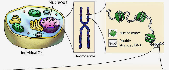
The human genome is divided into 23 chromosomes pairs, found in the nucleus of every cell. To keep these long linear polymers of DNA from getting all tangled up, the DNA of each chromosome is packaged into repeating structural units called nucleosomes. The DNA exists as a double–stranded structure with two twisting backbones running in opposite directions and four different bases: adenosine (A), thymine (T), guanine (G), and cytosine (C).
The visual representation of DNA above is completely interactive and can be rotated in 3-dimensions by clicking and dragging with your mouse.
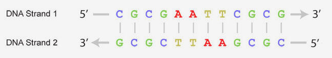
The four types of bases that make up DNA can form base-pairs between the two strands of double-stranded DNA. Adenosine only pairs with thymine and guanine only pairs with cytosine, making the two strands of DNA complimentary. The sequence of bases is often represented with abbreviated letters as shown below. It is this order of DNA bases that contains the key information needed for creating proteins and passing along genetic information.
For more information on DNA structure, explore the RCSB PDB's Molecule of the Month feature on DNA article by the molecular illustrator, David Goodsell.
What is a Genome?
The complete genetic information (DNA) that functions as the blueprint for creating an organism is called a genome. Each cell of the organism contains this genome. As the cells divide the genomic information is carried forward through generations.
The human genome is composed of 3.2 billion basepairs. In a heroic effort starting in the 1990s, several international research groups participated in sequencing the complete DNA sequence of the human genome. While the sequencing was completed in 2003, scientists are still working on reading and understanding the meaning and complete implications of the genomic sequence. Today it is possible to sequence an individual’s genome and figure out if they are genetically pre-disposed to specific diseases or how they will respond to certain treatments for a specific disease.
The Flow of Genetic Information - From Genes (DNA) to Protein
While double–stranded DNA has become one of the most iconic structures in modern biology, it is only part of the story. DNA is important in that it contains the information cells need to make proteins.
Proteins are made of smaller building blocks called amino acids that are linked together to form long chains. These protein chains spontaneously fold up into compact 3-dimensional shapes following basic principles of chemistry and physics. Each type of protein has a unique sequence of amino acids, which determines its unique 3-dimensional shape and function.

The sequence of amino acids in a protein is encoded by the sequence of bases in the gene (DNA). (Read this over – – and over again – – and think about it until it makes sense to you). When a gene is "expressed" – or made into a protein – it is first copied into messenger RNA (mRNA) by the process known as transcription. RNA polymerase is an enzyme that synthesizes mRNA by reading your DNA. Messenger RNA is complementary to the template strand of DNA – following the base pairing rules (A pairs with U; G pairs with C). The short animation below shows transcription.
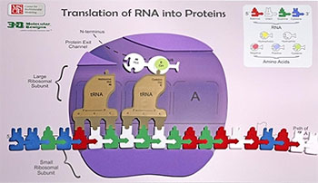
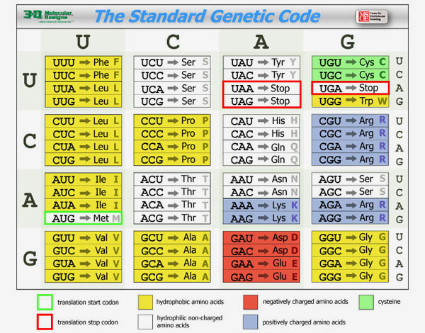
Once the mRNA has been created from the DNA, it is bound by a large macromolecular complex called a ribosome that reads this sequence of mRNA bases and builds a protein with a specific sequence of amino acids based on the mRNA sequence. The mRNA is read three bases at a time based on the code that is illustrated in The Standard Genetic Code shown below. Note that the code is degenerate – meaning that most amino acids are encoded by more than one three-base sequence (called a codon).
Special molecules called tRNA read the sequence of mRNA three bases at a time and add the correct amino acid to the growing protein chain based on the mRNA sequence. The short animation below shows this important process called translation.
Genomes and Personalize Medicine

Personal Genome Analysis
This excerpt from the 2013 HudsonAlpha Biotechnology Guide Book provides an overview of personal genome analysis, covering recent technological advances in the feild and how genome data is used.
Personalized Medicine
This excerpt from the 2013 HudsonAlpha Biotechnology Guide Book provides an overview of personalized medicine, describing how genome sequencing is being used by doctors to provide unique and individualized medical care to patients.

Diagnostic Clinical Genome and Exome Sequencing
This article from the New England Journal of Medicine discusses using genome and exome sequencing to diagnose clinical disorders.
This Year's Proteins
The table below lists the proteins that will be featured as the 2016 pre-build as well as the various on-site builds at invitational, regional, state and national competitions.
Be Sure to Click on Each Protein Listed Below to reveal additional resources that you should explore as you prepare for each level of competition. In addition to providing the pdb file for the protein that will be featured, these links also provide a copy of the original research paper (the "primary citation") that reported the protein's structure. Questions regarding the paper and the structure presented in the PDB file will be asked on the exam you will complete at each competition.
Pre-Build Model Sepiapterin Reductase 1sep.pdbCoordinates for the Model
The 2016 Pre-Build Model should represent amino acids 93-261 of chain A of the sepiapterin reductase protein based on the PDB file 1sep.pdb.
Background Information
An annotated version of the "primary citation" (the original research paper that reported this structure) is provided below. This is a very detailed research paper. You are not expected to read and understand the entire paper. To guide your reading, we have highlighted those sections of the paper that will provide you with a general introduction the structure and function of the protein.
Paper Abstract:
Sepiapterin reductase catalyses the last steps in the biosynthesis of tetrahydrobiopterin, the essential cofactor of aromatic amino acid hydroxylases and nitricoxide synthases. We have determined the crystal structure of mouse sepiapterin reductase by multiple isomorphous replacement at a resolution of 1.25 angstrom inits ternary complex with oxaloacetate and NADP. Thehomodimeric structure reveals a single-domain a/bfold with a central four-helix bundle connecting two seven-stranded parallel b-sheets, each sandwiched between two arrays of three helices. Ternary complexes with the substrate sepiapterin or the product tetrahydrobiopterin were studied. Each subunit contains a specific aspartate anchor (Asp258) for pterin-substrates, which positions the substrate side chain C19-carbonyl group near Tyr171 OH and NADP C49N. The catalytic mechanism of SR appears to consist of a NADPH-dependent proton transfer from Tyr171 to the substrate C19 and C29 carbonyl functions accompanied by stereospecific side chain isomerization. Complex structures with the inhibitor N-acetyl serotonin show the indoleamine bound such that both reductase and isomerase activity for pterins is inhibited, but reaction with a variety of carbonyl compounds is possible. The complex structure with N-acetyl serotonin suggests the possibility for a highly specific feedback regulatory mechanism between the formation of indoleamines and pteridines in vivo.
Note that your pre-build model is based on the structure of a mouse sepiapterin reductase protein, as described by 1sep.pdb. We chose the mouse enzyme for this protein modeling event because:
- It was the first structure of this protein that was solved (in 1999).
- Tetrahydrobiopterin was bound in the active site of this enzyme.
- The mouse enzyme is 74% identical (and 88% similar) to the human enzyme.
Additional Resources
Molecule of the Month - Tetrahydrobiopterin Biosynthesis
The molecular illustrator David Goodsell writes a monthly article for the RCSB Protein Databank called The Molecule of the Month. The August 2015 edition covers tetrahydrobiopterin biosynthesis and provides an excellent overview of sepiapterin reductase and tetrahydrobiopterin.
Coordinates for the Model
The 2016 Invitational On-site Model will be taken from a G-protein alpha subunit based on the PDB file 1gia.pdb.
Background Information
An annotated version of the "primary citation" (the original research paper that reported this structure) is provided below. This is a very detailed research paper. You are not expected to read and understand the entire paper. To guide your reading, we have highlighted those sections of the paper that will provide you with a general introduction the structure and function of the protein.
Paper Abstract:
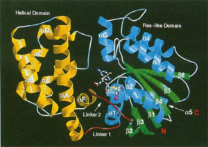
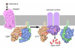
Mechanisms of guanosine triphosphate (GTP) hydrolysis by members of the G protein a subunit-p21 ras superfamily of guanosine triphosphatases have been studied extensively but have not been well understood. High-resolution x-ray structures of the GTPyS and GDP.AIF4- complexes formed by the G protein Giai demonstrate specific roles in transition- state stabilization for two highly conserved residues. Glutamine204 (Gin61 in p21 ras) stabilizes and orients the hydrolytic water in the trigonal-bipyramidal transition state. Arginine 178 stabilizes the negative charge at the equatorial oxygen atoms of the pentacoordinate phosphate intermediate. Conserved only in the Ga family, this residue may account for the higher hydrolytic rate of Ga proteins relative to those of the p21 ras family members. The fold of Gja1 differs from that of the homologous Gta subunit in the conformation of a helix-loop sequence located in the a-helical domain that is characteristic of these proteins; this site may participate in effector binding. The amino-terminal 33 residues are disordered in GTPyS-Gjai1, suggesting a mechanism that may promote release of the Ay subunit complex when the a subunit is activated by GTP.
Additional Resources
Molecule of the Month - G-Proteins
The molecular illustrator David Goodsell writes a monthly article for the RCSB Protein Databank called The Molecule of the Month. The October 2004 edition covers G-Proteins and provides an excellent overview of G-Proteins and the role they play in neurotransmission.
Coordinates for the Model
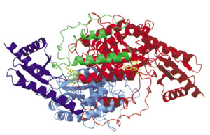
The 2016 Regional On-site Model will be taken from the DOPA decarboxylase protein based on the PDB file 1js3.pdb.
Background Information
An annotated version of the "primary citation" (the original research paper that reported this structure) is provided below. This is a very detailed research paper. You are not expected to read and understand the entire paper. To guide your reading, we have highlighted those sections of the paper that will provide you with a general introduction the structure and function of the protein.
Paper Abstract:
DOPA decarboxylase (DDC) is responsible for the synthesis of the key neurotransmitters dopamine and serotonin via decarboxylation of L-3,4-dihydroxyphenylalanine (L-DOPA) and L-5-hydroxytryptophan, respectively. DDC has been implicated in a number of clinic disorders, including Parkinson’s disease and hypertension. Peripheral inhibitors of DDC are currently used to treat these diseases. We present the crystal structures of ligand-free DDC and its complex with the anti-Parkinson drug carbiDOPA. The inhibitor is bound to the enzyme by forming a hydrazone linkage with the cofactor, and its catechol ring is deeply buried in the active site cleft. The structures provide the molecular basis for the development of new inhibitors of DDC with better pharmacological characteristics.
In the video below,Tim Herman, Ph.D. from the MSOE Center for BioMolecular Modeling discusses the primary citation for the regional onsite model for the 2016 Science Olympiad Protein Modeling Event.
Coordinates for the Model
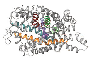
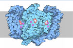
The 2016 State On-site Model will be taken from the dopamine transporter protein based on the PDB file 4m48.pdb.
Background Information
A copy of the "primary citation" (the original research paper that reported this structure) is provided below, which will provide you with a general introduction to the structure and function of the protein.
Paper Abstract:
Antidepressants targeting Na+/Cl- coupled neurotransmitter uptake define a key therapeutic strategy to treat clinical depression and neuropathic pain. However, identifying the molecular interactions that underlie the pharmacological activity of these transport inhibitors, and thus the mechanism by which the inhibitors lead to increased synaptic neurotransmitter levels, has proven elusive. Here we present the crystal structure of the Drosophila melanogaster dopamine transporter at 3.0 angstrom resolution bound to the tricyclic antidepressant nortriptyline.The transporter is locked in an outward open conformation with nortriptyline wedged between transmembrane helices 1, 3,6 and 8, blocking the transporter from binding substrate and from isomerizing to an inward-facing conformation. Although the overall structure of the dopamine transporter is similar to that of its prokaryotic relative LeuT, there are multiple distinctions, including a kink in transmembrane helix 12 halfway across the membrane bilayer, a latch-like carboxy-terminal helix that caps the cytoplasmic gate, and a cholesterol molecule wedged within a groove formed by transmembrane helices 1a, 5 and 7. Taken together, the dopamine transporter structure reveals the molecular basis for antidepressant action on sodium-coupled neurotransmitter symporters and elucidates critical elements of eukaryotic transporter structure and modulation by lipids, thus expanding our understanding of the mechanism and regulation of neurotransmitter uptake at chemical synapses.
Additional Resources
Molecule of the Month - Neurotransmitter Transporters
The molecular illustrator David Goodsell writes a monthly article for the RCSB Protein Databank called The Molecule of the Month. The March 2014 edition covers neurotransmitter transporters and provides an excellent overview of the dopamine transporter.
Coordinates for the Model
The 2016 National On-site Model will be taken from the Dopamine Receptor based on the PDB file 3pbl.pdb.
Background Information
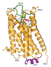
A copy of the "primary citation" (the original research paper that reported this structure) is provided below, which will provide you with a general introduction to the structure and function of the protein.
Paper Abstract:
Dopamine modulates movement, cognition, and emotion through activation of dopamine G protein–coupled receptors in the brain. The crystal structure of the human dopamine D3 receptor (D3R) in complex with the small molecule D2R/D3R-specific antagonist eticlopride reveals important features of the ligand binding pocket and extracellular loops. On the intracellular side of the receptor, a locked conformation of the ionic lock and two distinctly different conformations of intracellular loop 2 are observed. Docking of R-22, a D3R-selective antagonist, reveals an extracellular extension of the eticlopride binding site that comprises a second binding pocket for the aryl amide of R-22, which differs between the highly homologous D2R and D3R. This difference provides direction to the design of D3R-selective agents for treating drug abuse and other neuropsychiatric indications.